
Figure 32
Pages 22-41
The Expanded Concept of Tree Decay
The expanded concept of tree decay has four major parts:
1) WOUNDS start the PROCESSES (fig. 32)
Figure 32
2) MANY ORGANISMS are associated in the processes SUCCESSIONS of 4 organisms are involved in the infection of the wood. (fig. 33)

Figure 33
3) The tree REACTS to the wound. The living cells behind the wound
react immediately. (fig. 34)

Figure 34
The COMPARTMENTED tree COMPARTMENTALIZES the injured and infected tissues. (fig. 35)

Figure 35
4) DISCOLORED and DECAYED wood results, but this wood is COMPARTMENTALIZED by WALLS 1,2,3, and 4. (fig. 36)

Figure 36
Click here for more information on color codes of fig. 35 & 36.
Pages 22-23
Here Are Some Basic Patterns of COMPARTMENTALIZATION and SUCCESSIONS After One Wounding Period.
COLOR CODES
Red-Tree response (chemical protective reactions).
Green-Position of pioneer microorganisms (can be bacteria, decay fungi,
or nondecay fungi). Wood in this area is usually discolored; its cell contents
are altered.
Brown-Position of decayed wood; cell walls are digested.
Five branch stubs. Strong compartmentalization. The wound closure is complete. (fig. 37)

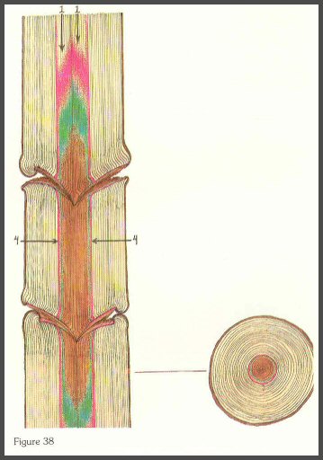
(Below) Four large open stubs. Diameter of the tree at time of branch death is the diameter of the column of decayed wood. Discolored wood surrounds the decay. Pioneer organisms are in green zone. (fig. 38)
(Below) Closed minor wound after 5 years. Invasion is well compartmentalized. (fig. 39)

(Below) Two large open wounds after 10 years. Column of discolored and decayed wood is diameter of tree when wounded. This pattern will be the same in trees that have heartwood. INJURED AND INFECTED TISSUES IN HEARTWOOD ARE ALSO COMPARTMENTALIZED. (fig. 40)

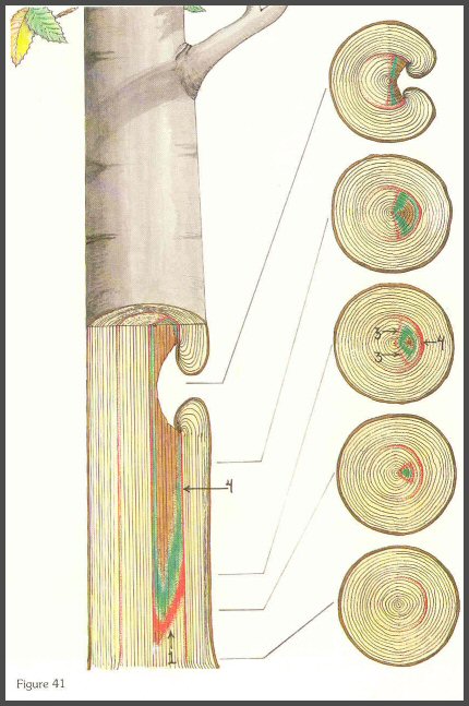
(Below) One severe wound after 10 years. The discolored and decayed wood is confined to one side of the tree. Although the red zone is shown on the side opposite the wound, no anatomical changes will be seen. All the wood within the entire cylinder present at the time of wounding will be slightly altered; in most cases, this will be too subtle to be visible. (fig. 41)
Pages 24-25
WOUNDS start the processes that could lead to discolored and decayed wood.
The classical concept and the expanded concept both recognize wounds as the
starting point for the processes. Trunk wounds can be caused by a variety
of agents: insects, birds, small and large animals, wind, ice, snow, temperature
extremes, chemicals, and people and some of their activities. Often the
wound is seen but not the agent that inflicted it, such as the porcupine wound
in this pine. (fig. 42)

And the black bear wound on the western hemlock. Wall 4 forms as a cylinder and the decay develops as a cone within the cylinder as Walls 1, 2, and 3 give way to microorganisms. (fig. 43)

Pages 26-27
Root and butt decay associated with wounds are also compartmentalized in gymnosperms
(fig. 44) and angiosperms. (fig. 45)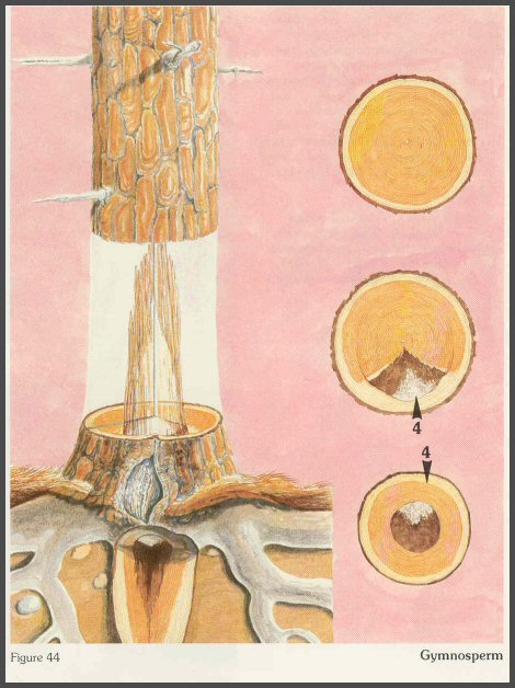

Figure 45
Angiosperm
Ganoderma applanatum
Pages 28-29
WOUNDS are of two major types:
1) BRANCH-The branch wounds expose all tissues from the new outer tissues to
that of the older tissues in the center of the tree. (fig. 46)

2) OUTER CORE-The outer core wounds usually expose only the most recently formed tissue. This depends of course on the depth of the wound. (fig. 47)

When microorganisms infect, they grow from compartment to compartment. The column of discolored or decayed wood that is seen macroscopically will be a composite of many columns as seen in these radial sections through drill wounds. (fig. 48)

In a vertical plane, each growth ring ("tree") will have its own column of discolored and decayed wood. The response of the "older trees" or innermost rings is slower and weaker than that of the "younger trees:' (fig. 49)

The "holding strength" of Wall 1 decreases as the injury goes inward. The older "trees" have a weaker response. This action determines the shape of the column, shown here from a radial view. (fig. 50)

In a sense, the wounds set the stage. The DISCOLORATION PROCESSES begin immediately after wounding when the tree reacts-both by chemical reaction and by plugging. When the pioneer microorganisms invade, the discoloration processes may intensify. The DECAY PROCESSES begin when microorganisms begin to digest cell walls. Many factors affect the rate of the discoloration and decay processes- severity of the wound, position, size, time of year of wounding, wounding agents that import materials into the wound, and the types of microorganisms that infect.
Pages 30-31
A branch wound will usually have this shape of column. Of course, there are many variations to this shape. (fig. 51)

Figure 51
Page 32
Ring rot is one configuration associated with branch infections. (fig. 52)

Figure 52 Fomes pini
Page 33
When many branches die at about the same time, the entire central column of the trunk may be decayed. The diameter of the column will be the diameter of the tree when the branches died. Although the death of the branches determines the diameter of the internal column, the fungi associated with the decay in the column may have entered the branch and trunk after a long growth period in the dying branch. Some fungi, such as Echinodontium tinctorium and possibly Fomes pini and others, first infect minute branchlet stubs on the living branch and therefore usually do not infect the large, freshly exposed branch stubs. (fig. 53)

Branches are often decayed by a wide variety of microorganisms that do not enter the trunk. Branch decay of this type is very beneficial as the tree is pruned when the branches fall. (fig. 54)

Pages 34-35
An outer core wound will usually have this shape, but again, many variations exist. In nonheartwood- forming trees, the column will be as shown. In heartwood-forming trees, the column will extend farther along the heartwood-sapwood boundary that was present at the time of wounding.
Also, concentrations of pigments and oxidized protective materials will usually be greater along the side of the column closest to the cambium. The column stays to the pith side of Wall 4. (fig. 55)

Page 36
Some Typical Patterns of Discolored and Decayed Wood Associated with Wounds
Near the wound, the individual columns within each growth ring will be
clustered. Some of the various shapes of discolored and decayed wood will
be seen as on transverse wedge-shaped sections in the next series of diagrams.
(fig. 56)

- - - - - - - - - - - -
(Below 3 Figures) CODES FOR ALL DIAGRAMS
Green-Discolored wood
Brown-Decayed wood
Orange-Wall 4
- - - - - - - - - - -
The eye sees a macroscopic view like this. (fig. 57)

Figure 57
But the mind's eye SHOULD see a diagrammatic view like this. Keep this
in mind while viewing the following diagrams. (fig. 58)

Figure 58
Walls 2, 3, and 4 are pointed out by arrows in this drawing. These walls will be present in other diagrams, but no arrows will be shown. (fig. 59)

Figure 59
Page 37
Six-Year-Old Severe Type Wound
The discolored wood forms the typical triangular pattern into the pith. (fig.
60)

Four-Year-Old Moderate Type Wound
The compartments directly beneath the wound were the only ones affected (fig.
61)

Multiple Wounds: A central 10-year- old wound associated with a central column of discolored wood. A later 4- year-old wound with a triangular column of discolored wood that developed to the boundary of the inner barrier zone surrounding the 10- year-old wound. (fig. 62)

Multiple Wounds: A central 10-year- old wound with a central column of discolored wood. A 2-year-old wound with a small column of discolored wood did not spread into the older, more central column. (fig. 63)

Page 38
A Deep Drill Wound: The tissues between the end of the drill wound and the inner column of discolored wood will discolor slightly. The radial walls (Walls 3) limit the lateral spread of the discolored wood. (fig. 64)

When the drill hole is shallow, the compartments between the inner column and the tip of the drill hole may remain healthy or nondiscolored. This is the case when shallow injection wounds are inflicted. (fig. 65)

(Below) A slanted drill hole usually gives this type of pattern. Note that the discolored compartment between the central column of discolored wood and the tip of the drill hole follow the ray pattern inward (Wall 3). The column beyond the end of the drill hole does not continue in the same direction as the hole. Often the side of the hole closest to the cambium will be darker from an accumulation of phenols. (fig. 66)

Page 39
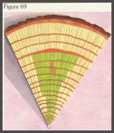
A series of seven sections from above a wound similar to that shown on page 39 figure 56. Each section is approximately 20 centimeters above the other. Note the pattern of discolored and decayed wood and the position of the barrier zone. (figs. 67-73)


Page 40
(Below) Four sections showing the pattern of discolored and decayed wood associated with multiple wounds on four trees. A multitude of specific multiple patterns is possible depending on the wounds inflicted, their severity, position, and time between woundings. (figs. 74-77)


Page 41