Note from Web Master (Often the word callus is used in this doc when the actual material is woundwood. You can look up the two words at www.treedictionary.com)
322

Figure 1. Drill wounds in red maple 3 months after treatment 1 ml of Ethrel, left and control, right. The small depressions show where the chips of wood were extracted for isolation of microorganisms. This pattern of isolations was repeated on many other wounds. Six wood chips were placed into agar in one Petri dish.

Figure 2. Sporophores of Panus stipticus on wounds treated with Ethrel after 3 years.

Figure 3. Decayed wood associated with wounds treated with Ethrel (A and B). The decayed wood supported sporophores of Panus stipticus.

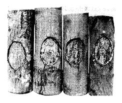
Figure 4. Thick bands of callus associated with 2-year-old control wounds on one red maple. There was very little cambial dieback.

Figure 5. Thin bands of callus associated with 2-year-old control wounds on one red maple. Large area of cambial dieback were associated with the wounds.

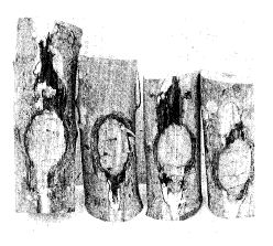
Figure 6. Thick bands of callus associated with 2-year-old wounds on one red maple treated with rosin acids (three wounds on right), and control (wound on left).
 Figure 7. Thin bands
of callus and extensive areas of cambial dieback associated with 2-year-old
wounds on one red maple treated with rosin acids (three wounds on right) and
control (wound on left).

 Figure 9.
Large areas of cambial dieback associated with 2-year-old wounds on one red
maple treated with rosin acids (three wounds on right) and the control (wound on
lefty. Bark peeled to show extent of dieback.