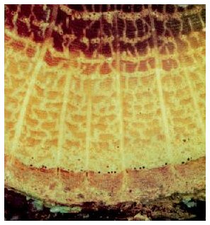
 WHITE
OAK, Quercus alba
 By Dr. Alex Shigo
 WHITE
OAK, Quercus alba
 The growth rate of this tree was decreasing for the last 15 years.
The last seven growth increments consisted of only earlywood vessels. The
tree was a victim of repeated attacks the last seven years by gypsy moths,
Lymantria dispar. (I was watching this tree for many years until it was
cut for a housing development.)
Trees keep a very accurate log of all events that affect their lives The log is kept in the wood, and to read that log you must under stand the simple language of tree anatomy. Trees respond to the ever changing environment, and to injuries and infections. Because pruning and removing trees are major activities of most tree companies. arborists have many opportunities to autopsy a tree and read its log.
Autopsy, which comes from the Greek word autopsia, means to see
for yourself. It is often mistaken for the word for necropsy, which means
the study of the dead. The usefulness of an autopsy depends on knowing
where to look, what to look for, and the meaning of what you see. You must
be able to see details fast. We have a special name for people who can
see fast. We call them lucky!
Here is check list for some major features to look for and record
when reading the tree's log.
1. Growth increments - tree age, patterns of wide or narrow increments,
eccentric growth patterns, date when increments began to increase or decrease
in width, colors.
2. Wood type - diffuse-porous, semi-ring porous, ring porous, conifer
resinous, conifer non-resinous, tropical, monocot.
3. Energy reserves, I²,KI to determine amount of starch and volume of
wood with starch.
4. Wound history - date of wounds to the year, or, in some cases, the
week they were inflicted.
5. Branch history - when branches died. If pruned, how they were pruned
and the defect associated with the branches.
6. Cracks - boundaries from wounds, ring shakes, radial cracks, cracks
in bark only, cracks in wood and bark, wetwood in cracks.
7. Animal wounds - bird peck wounds and ring shakes, squirrel wounds,
other animal wounds.
8. Closure patterns of wounds - ram's horns, cracks, woundwood ribs,
discolored wood associated with cracks.
9. Discolored wood and wetwood - patterns of infected wood, CODIT walls,
callus, odors, internal checking patterns.
10. Decayed wood - white rot, brown rot, zone lines in rotted wood,
CODIT walls, sporophores.
11. Resin ducts - traumatic ducts in non-resinous conifers.
12. Tyloses - traumatic tyloses in wood that does not normally form
them.
13. Insects - galleries, in bark, in bark and wood, insect, ants, termites;
galleries clean or full of frass.
14. Reaction wood - compression wood in conifers. (You cannot see tension
wood.)
15. Injection and implant wounds - separate cloumns of discolored wood,
or columns coalescing.
As you learn more about tree anatomy, many other details of the tree's
log will become obvious.
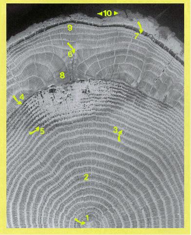 RED OAK,
Quercus rubra, 38 years old, with a closed and compartmentalized
wound.
1. A star-shaped pith as with most oak species. There is no pith in roots. Note the five-lobed growth increments near the center of the tree.
2. Red oak is a ring-porous tree that forms large vessels first in the growing period and smaller vessels later. Wide and narrow rays radiate from the center of the tree. Oaks have a darkly colored protection wood called heartwood. All cells are dead in the heartwood.
3. Some events caused the tree to start decreasing its growth rate at this time. Note the decreasing width of the growth increments.
4. The tree was wounded by buckshot during a dormant period nine years before it was cut. The barrier zone boundary between the growth increments indicates a wound during the dormant period. A wound during the growing period will cause a barrier zone boundary to form within the growth increment.
5. A dark boundary called a reaction zone borders the column of decayed wood in the heartwood. Note that the boundary is darker in color than the heartwood, but as decay develops, the darker wood changes to a lighter color. This is called white rot, because the cellulose and lignin are digested by the fungi.
6. The woundwood ribs closed the wound in five years. Note the bark between the ribs of woundwood
7. The tree was cut just as the first vessels were forming. Since
vessels begin to form as the leaves are expanding, this tree was cut around
the second week of May in New Hampshire.
8. The size of the woundwood ribs were very large before the wound was closed. Note that the woundwood ribs contained mostly dense wood with few vessels.
9. After wound closure, the size of the growth increments were about the same as those before the tree was wounded.
10. The pointer to the left shows new bark with a smooth corky
layer-phellem. The pointer to the right shows the old original rough phellem
of the tree.
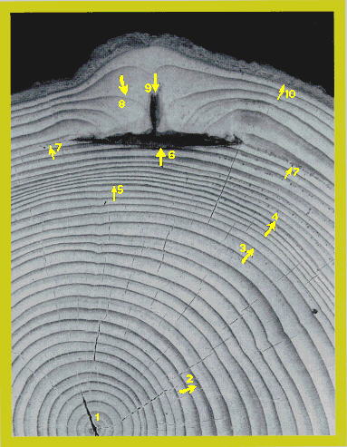 RED SPRUCE,
Picea nibra, 40 years old, with a closed and compartmentalized wound.
Spruce trees are conifers that have tracheids, not vessels, for water transport
and mechanical support. As the growth increment increases, the tracheids
that formed later have thicker walls. These are called fiber tracheids.
1. The tree started to grow at an even rate. After six years, it began to lean slightly to the left.
2. After 13 years, it began to lean slightly to the right. Note the larger and darker bands of compression wood.
3. At this time, the tree was injured. Note the sudden decrease in growth rate.
4. Spruce trees have very few resin ducts in healthy wood. The wood is a non-resinous type. However, when the tree is injured, resin ducts, called traumatic ducts, often form. The ducts appear as dark spots in the wood. Even though the wound is not shown in the photo, you can be sure there was a wound nearby on the tree.
5. A very narrow ring shake or separation indicates a small wound near where this specimen was cut. Note also the sudden decrease in growth rate to the left of the arrow.
6. The wound penetrated one growth increment. A very strong "CODIT wall 2" resisted deeper spread into the tree. Note the dark and of fiber tracheids at the arrow point. The wood was altered chemically as a protection wood after wounding.
7. Note both arrows showing the barrier zone that formed after wounding. When trees are wounded during the growing period, it is possible to date the wound to within a week of when it was inflicted. The barrier zone is slightly beyond the middle of the growth increment indicating that the wound was inflicted about four to five weeks after new needles began to form. Under normal conditions, it takes about six to eight weeks after needle or leaf formation before the growth increment is fully formed. This tree was wounded around the third week of June in New Hampshire.
8. Thick woundwood ribs began to close the wound.
9. The wound was closed in four years.
1 0. The tree was cut about a week after new tracheids began
to form. It was cut the third week of May, and therefore the wound was
six years and about four weeks old.
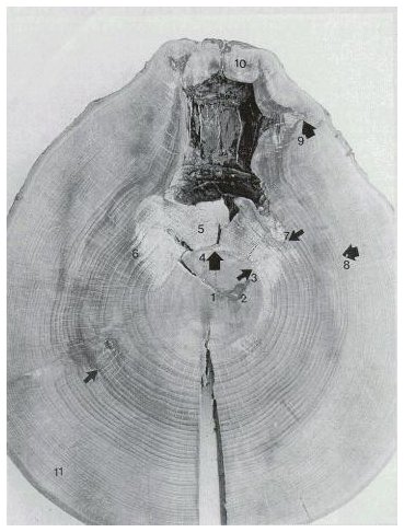 AMERICAN BEECH,
Fagus grandifolia, about 110 years old, with a column of compartmentalized,
decayed wood associated with an old, dead branch.
1. Beech has diffuse-porous wood. All vessels are about the same size and arranged evenly throughout the growth increment. Beech will tolerate low light and often it will grow very slowly, as shown here, as an understory tree. When it is released into light, it grows rapidly.
2. A small wound with decay was compartmentalized in the center of the tree.
3. The tree lost branches at this time and a core of protection wood formed. This type of protection wood is called false heartwood, because the death of branches triggers the process. Heartwood formation in oaks is a genetically controlled aging process. The false heartwood, like heartwood, resists the spread of decay.
4. Note that the decay associated with the dead branch did not spread into the column of false heartwood. It could be that the events that brought about the formation of false heartwood--dying branches--also released the tree into more light. Note that after a few years, the growth increments increased greatly in width.
5. The cellulose and lignin were both being digested, indicating a white rot. The fungi did not grow into the central core of dense, slow growing, false heartwood.
6. Note that the decay spreads first into the earlywood of each growth increment. This pattern results in a tooth-like margin to the column of defect when viewed in cross section.
7. The decay associated with the branch did not spread outward into the column of discolored wood.
8. The limits for the column of discolored wood were set by the cracks that formed as the woundwood closed the wound.
9. A crack where the woundwood formed about the dead branch.
1 0. The curling woundwood ribs formed ram's horns. The cracks
that form in this way often extend the columns of discolored and decayed
wood beyond the barrier zone and wood present at the time of branch death
or of wounding.
 Dr. Alex L.
Shigo doing an autopsy.
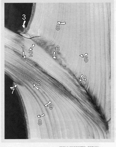 CANADIAN HEMLOCK,
Tsuga canadensis, about 40 years old, with a dead branch.
1. The pith of the branch was infected and darkly discolored.
2. Both arrows show the branch protection zone that formed after the branch died.
3. The branch bark ridge.
4. Note the invaginated increments indicating included bark to this point. Note also the dark color of the wood from the arrow downward toward the trunk. This indicates that the increments were squeezing together to the point of cell death.
5. For some reason, the increments began to form normally at this point. A much narrower growth increment formed at this time, suggesting a possible injury.
6. A different type of checking pattern can be seen where the normal branch-trunk collars began to form. Compare area 6 to area 4.
7. A crack formed after the branch died. The branch died about 12 years before the tree was cut.
8. There was a sudden decrease in growth rate at this time. Note both arrows 8. The number of growth increments above the branch are equal to those below the branch from arrows 8 to the bark.
9. Compression wood began to form here.
“An author, lecturer and consultant, Dr. Shigo started Shigo and Trees, Associates twenty years ago after retirement from the U.S. Forest Service.”
Reproduced with permission of Tree Care Industry and Dr. Alex L. Shigo.
The article was published in Volume VII, Number 6 -June 1996 of TCI.
This site is dedicated to the remembrance of Robert Felix who for many years worked very hard for the improvement of the tree care industry: 1934-1996.
Dictionary MAIN
PAGE
Text & Graphics Copyright © 2009
Keslick & Son Modern Arboriculture
Please report web site problems, comments and words of interest,
not found.
Contact